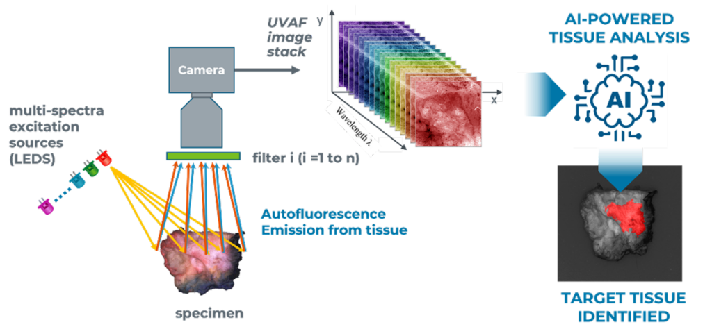
OncoLux’s’ AURORA™ Multispectral Specimen Imaging Platform is designed for Target Tissue Identification through the detection
of Intrinsic Biomolecular Markers using Tissue Autofluorescence Mapping
STEP 1
Multi-Spectral Illumination
STEP 2
Multi-Spectral Detection
STEP 3
Train AI/ML Algorithms
Multispectral Autofluorescence Imaging
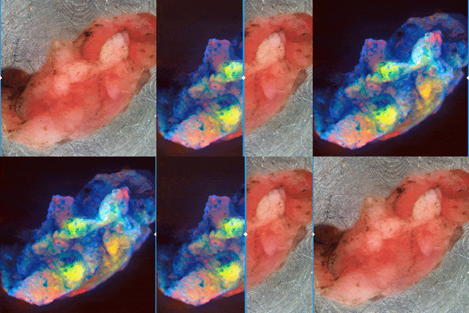
OncoLux’s AURORA™ Imaging allows the differentiation of tissue types & structures through differential autofluorescence and direct, diffuse reflectivity imaging
The example illustrates a TURBT specimen in RGB under a false-color three-channel autofluorescence composite image. Different colors and shades represent the different biomolecular constituents present in the tissue.
Tumor Margin Imaging
During cancer resection surgery, surgeons often cannot completely & definitively distinguish cancer from surrounding normal tissue. This can lead to incomplete tumor removal, unintended patient injury, re-operations, and/or higher likelihood of recurrence.
This is true in >20% of cancer surgeries, creating a multi-billion-dollar per year problem in the U.S. alone.
The OncoLux Solution leverages our MSAF enhanced tissue imaging technology to learn the fingerprint of malignant tissue to highlight regions of potential positive margins intraoperatively. This type of real-time imaging during surgery provides the surgeon with actional data to help improve patient outcomes, and improving the odds of preserving function and reducing disease recurrence.
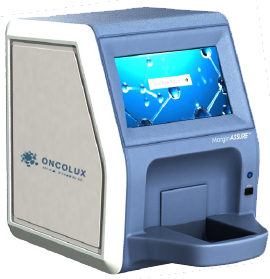
The OncoLux MarginASSURE™ Specimen Imager is a bench top unit based on our MSAF imaging technology platform, designed for cancer margin and residual cancer discrimination. Powered by our cancer classification AI-based algorithms OncoSight AI™, the technology has been demonstrated in breast and prostate cancer studies on hundreds of patient samples.
MarginASSUR™ uses AI-powered imaging to detect cancer margins and residual disease.

The OncoLux MarginASSURE™ Specimen Imager is a bench top unit based on Multi-Spectral Autofluorescence imaging technology, is designed for cancer margin and residual cancer discrimination and is powered by our tissue classification AI-based algorithms OncoSight AI™
Real-time imaging during prostatectomy could improve the odds of preservation of function and reduce recurrence for 90,000 men in the U.S. per year.
60% reduction in the breast cancer positive margin rate nationwide could avoid reoperations for 30,000 women annually.
Real-time bladder resection imaging could avoid 70,000 repeat TURBT procedures per year in the U.S.
PROBLEM
Surgeons cannot distinguish cancer from surrounding normal tissue, leading to incomplete tumor removal, unintended patient injury, re-operations and/or higher likelihood of recurrence. This is true in >20% of cancer surgeries creating > $1B per year problem in the U.S.
SOLUTION
The OncoLux Solution: Label free enhanced tissue imaging technology capable of highlighting regions of potential positive margins intraoperatively, providing surgeons actional data to help improve patient outcomes
TODAY
Surgeons often can’t definitively see the boundaries between healthy & cancerous tissue during tumor resection procedures
TOMORROW
Using Oncolux‘s enhanced imaging, surgeons will see and remove all cancer before closing.
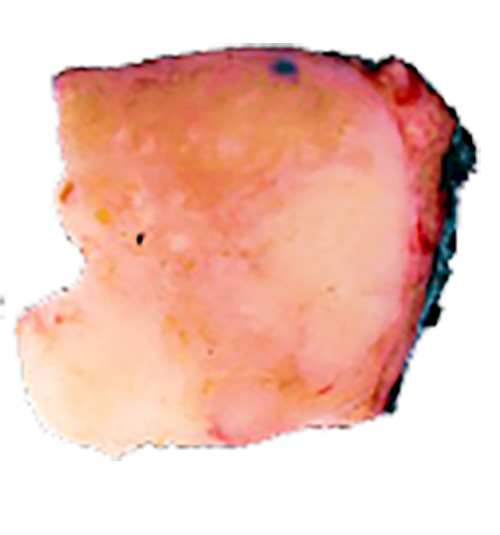
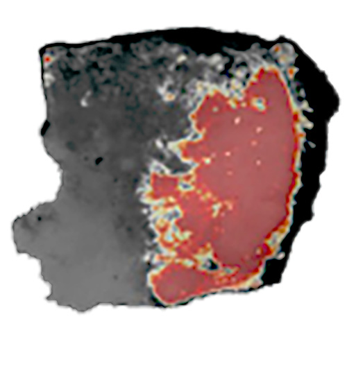
Slide left and right to compare tissue imaging.
We have demonstrated technology in breast bladder & prostate cancers:
Learn More about OncoLux’s Technology and Products
Breast
- 155 Patients
- 417 Specimens
- LOPO* Cross Validation
- 88% Accuracy¹

Bladder
- 98 Patients
- 173 Specimens
- LOPO Cross Validation
- 90% Accuracy²


Prostate
- 80 Patients
- 55 Specimens
- LOPO Cross Validation
- 86% Accuracy³

In general, surgeons attempt to balance clear margins with organ sparing and preservation of post-operative function and are demanding better intraoperative imaging tools to enable more precise surgeries and better outcomes.
