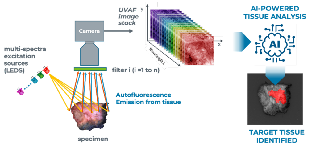
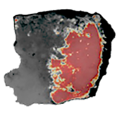
OncoLux Inc.

OncoLux’s’ AURORA™ Multispectral Specimen Imaging Platform is designed for Target Tissue Identification through the detection of Intrinsic Biomolecular Markers using Tissue Autofluorescence Mapping

STEP 1

Multi-Spectral Illumination

STEP 2

Multi-Spectral Detection

STEP 3

Train AI/ML Algorithms

Multispectral Autofluorescence Imaging

OncoLux’s AURORA Imaging  allows the differentiation of tissue types & structures through differential autofluorescence and direct, diffuse reflectivity imaging

The example illustrates a TURBT specimen in RGB under a false-color three-channel autofluorescence composite image. Different colors and shades represent the different biomolecular constituents present in the tissue.

AURORA’sAI-powered imaging distinguishes diseased from normal tissue.

The AURORA™ Specimen Imager is a bench top unit based on our Multi-Spectral Autofluorescence imaging technology, is designed for diseased and normal tissue discrimination and is powered by our tissue classification AI-based algorithms AURORAVision™

Units can be configured for high resolution, small specimen analysis: 10mm dia, and as such is suitable for biopsy specimen analysis. We have launched Aurora targeted for use in non-muscle invasive (NMI) bladder cancer and the analysis of specimens in transurethral resection of bladder tumor (TURBT) procedures.

Real-time imaging during surgery could improve the odds of preserving function and reducing recurrence.

MarginASSURuses AI-powered imaging to detect cancer margins and residual disease.

The OncoLux MarginASSURE™ Specimen Imager is a bench top unit based on Multi-Spectral Autofluorescence imaging technology, is designed for cancer margin and residual cancer discrimination and is powered by our tissue classification AI-based algorithms OncoSight AI

Slide left and right to compare tissue imaging. 

We have demonstrated technology in breast bladder & prostate cancers:

Learn More about OncoLux’s Technology and Products

Breast

  • 155 Patients
  • 417 Specimens
  • LOPO* Cross Validation
  • 88% Accuracy¹

Bladder

  • 98 Patients
  • 173 Specimens
  • LOPO Cross Validation
  • 90% Accuracy²

Prostate

  • 80 Patients
  • 55 Specimens
  • LOPO Cross Validation
  • 86% Accuracy³

In general, surgeons attempt to balance clear margins with organ sparing and preservation of post-operative function and are demanding better intraoperative imaging tools to enable more precise surgeries and better outcomes.